Spatial genomics is a true game-changer when it comes to studying the complexity of biological samples. Simply knowing what genes are expressed or the level of their expression is often not enough to gain a full understanding of biological systems. With spatial genomics, you get not only the genes that are expressed and the level, but also the POSITION within a cell or tissue and how they relate to each other. Truly a 3-dimensional view of biology.
Samples to Signals - full Spatial Service
Spatial genomics starts with tissue samples – FFPE or frozen – blocks or slides. Next Level Genomics can work with you to get your samples ready for success with spatial analysis with microtome services or from your own slides. Our histopathology and spatial experts will work with you to understand your project and needs. The selection of cell morphology markers, cell typing markers, and target panels can all be customized to maximize the data output for your project. Data analysis and visualization are available, or raw data can be delivered to you for your own analysis.
Let Next Level Genomics guide you on your spatial journey and discover the depth of data and discoveries available with the latest in spatial genomics technologies.
Context Is Everything
Why Spatial Genomics?
Spatial genomics is a true game-changer when it comes to studying the complexity of biological samples. Simply knowing what genes are expressed or the level of their expression is often not enough to gain a full understanding of biological systems. With spatial genomics, you get not only the genes that are expressed and the level, but also the POSITION within a cell or tissue and how they relate to each other. Truly a 3-dimensional view of biology.
Samples to Signals - full Spatial Service
Unlock the full potential of your research with our comprehensive spatial genomics solutions. At [Your Company Name], we transform biological samples into actionable insights by integrating advanced technologies and expert support throughout your project’s journey.
From Sample Preparation to Data Interpretation
•Project Consultation: Begin with in-depth discussions with our specialists to design a spatial genomics strategy tailored to your research objectives.
•Expert Sample Preparation: Our team excels in handling FFPE and frozen tissues, offering precision microtome sectioning to preserve the integrity of your samples.
•Advanced Staining Techniques: We provide a variety of staining options, including Hematoxylin and Eosin (H&E), immunofluorescence (IF), and custom protocols to highlight specific regions of interest.
•Cutting-Edge Spatial Profiling: Utilizing state-of-the-art platforms like GeoMx™ and CosMx™, we deliver high-resolution spatial transcriptomics and proteomics data, capturing the complexity of biological systems.
•Data Analysis and Validation: Our bioinformatics experts offer thorough data analysis and validation services, ensuring you gain meaningful insights from complex spatial data sets.
Spatial Proteomics
Method of the Year 2024
Spatial proteomics was recognized by Nature Methods as the Method of the Year 2024 for its transformative role in mapping proteins directly within intact tissues . Unlike transcriptomics, spatial proteomics captures the functional drivers of biology—proteins and their post-translational modifications—while preserving spatial context.
Technologies such as cyclic immunofluorescence (cycIF), CODEX, IBEX, MIBI, imaging mass cytometry (IMC), and emerging methods like deep visual proteomics (DVP) are generating highly multiplexed protein maps at cellular resolution. These advances are powering atlas-scale efforts such as HuBMAP and HTAN, accelerating discoveries in oncology, immunology, neuroscience, and precision medicine.
At Next Level Genomics, we bring these innovations to Singapore with the GeoMx Discovery Proteome Atlas (DPA), the highest-plex antibody-based spatial proteomics assay, enabling researchers to interrogate 1,200+ proteins—including 130+ post-translational modifications—directly in FFPE samples.
In addition, through CosMx Spatial Molecular Imaging, we support same-cell proteogenomics, allowing researchers to study RNA and protein within the same tissue sections. This integrated approach delivers a multi-dimensional view of disease biology, enabling faster translation from discovery to clinical insight.
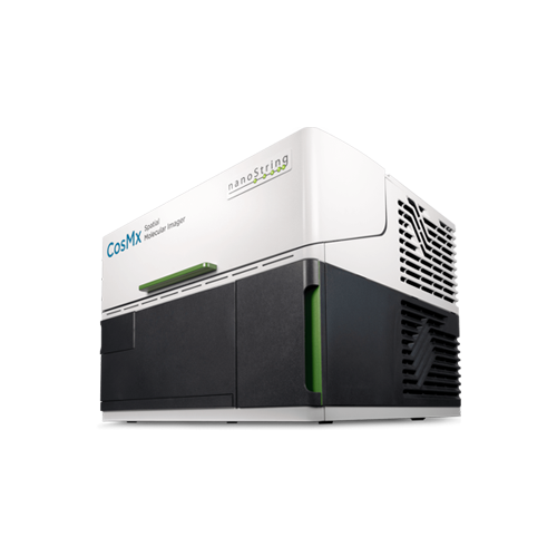
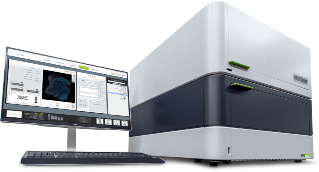
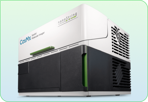
CosMx Spatial Molecular Imager (SMI)
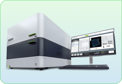
The CosMx Spatial Molecular Imager (SMI) from Bruker (formerly NanoString) is the highest-plex in situ analysis platform, enabling true spatial multiomics on both formalin-fixed paraffin-embedded (FFPE) and fresh frozen (FF) tissue at cellular and subcellular resolution.
CosMx SMI currently supports whole transcriptome (WTX) profiling as well as targeted RNA panels, together with validated protein detection. Researchers can rapidly quantify and visualize up to 6,000 RNA targets and 64 proteins within the same tissue section, with expanded protein + RNA assays in development.
This makes CosMx uniquely suited for proteogenomics, delivering an integrated view of RNA and protein biology in situ — accelerating discoveries in oncology, immunology, neuroscience, and translational research.
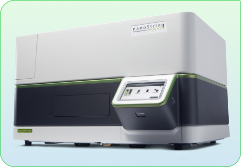
The GeoMx Digital Spatial Profiler (DSP) from Nanostring is a flexible, robust spatial multiomic platform for the analysis of FFPE and fresh frozen tissue sections. GeoMx is the only spatial biology platform that non-destructively profiles the expression of RNA and protein from distinct tissue compartments and cell populations with an automated and scalable workflow that integrates with standard histology staining.
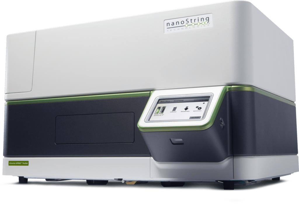
nCounter provides multiplexing up to 800 gene expression targets using direct detection technology. The workflow requires just 15 minutes of hands-on time for most panels using an automated benchtop processing system – requiring no amplification or technical replicates. Rapidly accelerate your biomarker validation and biomarker development with gene expression data you can count on.
The CosMx Spatial Molecular Imager from Nanostring is the highest-plex in situ analysis platform to provide spatial multiomics with formalin-fixed paraffin-embedded (FFPE) and fresh frozen (FF) tissue samples at cellular and subcellular resolution. CosMx SMI enables rapid quantification and visualization of up to 6,000 RNA and 64 validated protein analytes. [Soon to be whole transcriptome profiling].
Assays available on CosMx
Human 6K Discovery Panel RNA Assay
Human 1K Universal Cell Characterization RNA Assay
The GeoMx Digital Spatial Profiler (DSP) from Nanostring is a flexible, robust spatial multiomic platform for analysis of FFPE and fresh frozen tissue sections. GeoMx is the only spatial biology platform that non-destructively profiles expression of RNA and protein from distinct tissue compartments and cell populations with an automated and scalable workflow that integrates with standard histology staining.
nCounter provides multiplexing up to 800 gene expression targets using direct detection technology. The workflow requires just 15 minutes hands-on time for most panels using an automated benchtop processing system – requiring no amplification or technical replicates. Rapidly accelerate your biomarker validation and biomarker development with gene expression data you can count on.
Assays available on the nCounter
PanCancer IO 360
Breast Cancer 360 V2
PanCancer Immune Profiling
Tumor Signaling 360
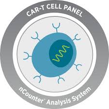
CAR-T Characterization
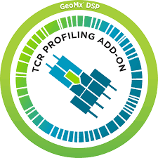
TCR Diversity
Direct Detection Technology
Gene Therapy Optimization Panel
No Sample Prep Needed – No CDNA Conversion, No RT, No Amplification
Gene Therapy Optimization Panel
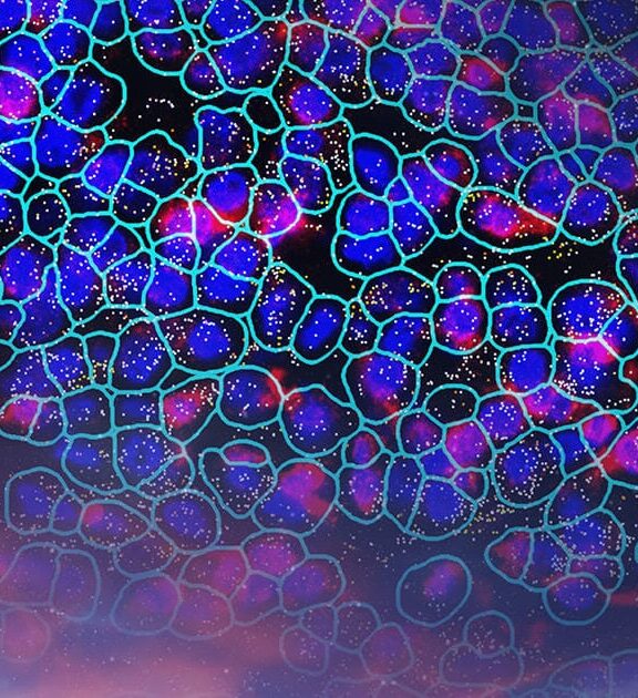
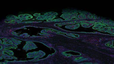
Sample Image of Data
Auxillary Spatial Services
Comprehensive Spatial Genomics Support: From Project Design to Data Validation
Our team provides end-to-end support for your spatial genomics projects, starting with in-depth project discussions to ensure optimal experimental design. We specialize in sample preparation, including expert FFPE-block and frozen tissue sectioning using microtome techniques. Additionally, we offer a wide range of staining options, including H&E, immunofluorescence (IF), and other custom staining protocols. Our services extend to validation, ensuring the accuracy and reproducibility of your spatial data. Let us partner with you throughout your spatial analysis workflow to deliver high-quality results.
We are experienced in FPPE-block and frozen tissue microtome slicing and slide preparation for spatial genomics analysis. H&E staining and other staining services are also available.
Spatially resolved architecture of organ tissues at the cellular and molecular level
Whole transcriptome data sets of organ tissues with spatial context
Contribute to regional and global consortia on cell organization and function
Biomarker Discovery
Identify and reliably measure biomarkers playing a critical role at every stage of the drug development process.
Generate robust exploratory biomarker data from limited and challenging sample types
Bulk gene expression analysis, whole transcriptome spatial analysis, or single cell and sub-cellular spatial analysis methodologies
Tumor Microenvironment Profiling
Understand tumor heterogeneity
Understand impact of the tumor microenvironment on the immune response
Characterize the microenvironment along the tumor invasive margin
Identify cellular neighborhoods that reveal tumor heterogeneity
Drug Mechanism of Action & Therapeutic Response
Measure treatment response in clinical trials
Discover and validate spatial biomarkers
Reveal functional changes within a tumor at single cell resolution
Molecular Subtyping of Disease and Disease Progression
Discover novel pathways and molecular disease subtypes using spatial profiling
Choose which regions to profile and segment each region into different compartments using fluorescent staining patterns as a mask to profile expression in certain tissue types or cell populations
Cell Atlas & Characterization
Spatially resolved architecture of organ tissues at the cellular and molecular level
Whole transcriptome data sets of organ tissues with spatial context
Contribute to regional and global consortia on cell organization and function
Applications of Spatial Genomics
Human 6K Gene Panel
6000 RNA targets – the most significant genes across every biological pathway + over 400 ligand-receptor pairs.
A nuclear stain plus four protein markers for cell segmentation.
Customize with up to 200 additional RNA targets.
Spatially analyze virtually the entire Reactome at the single-cell level.
Human 1K Gene Panel
Expression of 1000 highly curated targets at subcellular resolution.
Customize with up to 50 of your targets.
Up to five CosMx Segmentation Markers.
Provides robust tissue mapping, cell typing, and analysis of cell states and interactions on a wide range of tissues and solid tumors.
Human Immuno-oncology Panel
Expression of 100 transcripts covering immune cells, their activities, and aspects of tumor biology relevant to IO therapies.
Customize with up to ten of your own targets.
100-plex format offers faster turnaround and lower overall cost than high-plex panels.
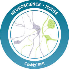
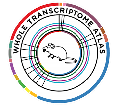
Mouse Neuroscience Panel
Robust cell typing, cell-cell interaction analysis, and more in the mouse brain and other neuronal tissues.
1000 highly curated targets at subcellular resolution and customize with up to 50 of your own targets.
Identification of 42 distinct cell types in the mouse brain.
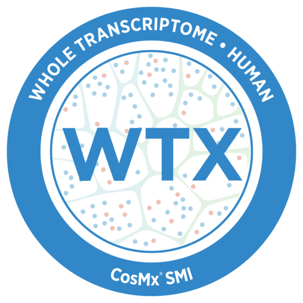
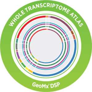
Whole Transcriptome Assay
Unbiased view of 18,000+ protein-coding genes – human or mouse.
Robust and sensitive performance on FFPE or Fresh Frozen samples.
Compatible with RNAscope® and antibody morphology markers.
For use with next-generation sequencer (NGS) readouts and compatible with DSP Data Center.
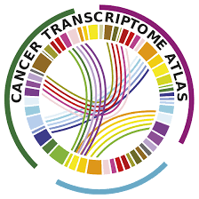
Cancer Transcriptome Atlas
Comprehensive profiling of tumor biology, the tumor microenvironment, and the immune response.
RNA expression of over 1,800 genes.
Measure clinically-derived gene sets, including the 18-gene Tumor Inflammation Signature (TIS), known to be associated with response to PD-1/PD-L1 inhibitor pathway blockade, and the 50-gene Prediction Analysis of Microarray 50 Signature (PAM50), known to be associated with breast cancer metastasis.
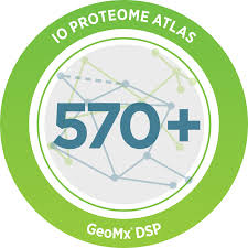
Immuno-oncology Proteome Atlas
Non-destructively profile over 570 proteins in different tissue compartments, such as the tumor, microenvironment, and immune infiltrate.
The highest-plex spatial proteomics panel is available for FFPE and fresh frozen tissue sections.
Customize with up to 40 additional protein targets of your choice.
Be the first to discover new multiomic biomarkers.
Tumor Signaling 360 – 760 genes covering the core pathways and processes of the tumor.
nCounter® Gene Therapy Optimization Panel – 800 genes covering processes known to impact gene therapy development and manufacturing.
PanCancer Pathways – 770 genes for essential cancer pathways.
and many more.
Custom Panels
The nCounter® platform offers the flexibility to tailor assay content to meet individual project needs with a triad of custom solutions.
Custom CodeSets, Elements™ TagSets, PlexSet™ Reagents, and Plus Content.
Frequently Asked Questions
How do I know if spatial genomics is right for my research?
Spatial genomics provides the most in-depth picture of the biology of a sample – from the spatial context in which cells and interactions are taking place to the extra-cellular microenvironment to morphological changes. Spatial is orders of magnitude more information than single-cell RNAseq or other methods of examining cell signaling and response. If you would like to know not just who the players are in your sample but where they are and how they interact, spatial genomics is for you.
Can I customize the RNA transcripts I’d like to investigate with spatial transcriptomics?
Yes, all assays on the Nanostring systems allow for customization with the addition of probes for your specific genes of interest. And any antibodies against your favorite protein target can be labeled and utilized for spatial analysis on these systems. For the nCounter, the whole custom panel design and validation are available. Contact our spatial specialist for information on gene lists and how to customize assays for your needs.
How long does it take to perform spatial analysis?
Spatial analysis involves slide preparation, processing, and scanning on the instruments. Slide preparation can be done by Next Level Genomics, or you can pass your own slides for processing. Slide processing takes 1-2 days, and they are ready to go onto the instrument. On the GeoMx DSP platform, scanning takes just a few hours, and regions of interest are selected and captured for downstream NGS analysis. NGS sequencing can take a few weeks before the data is available for final analysis. On the CosMx platform, the slides are scanned and fields of view are selected for analysis, and over 4–8 days, the data is collected and captured in real-time. Upon completion of the CosMx run, the data is available on the cloud-based AtoMx platform for immediate analysis.
How do I analyze my spatial data?
The Nanostring Spatial ecosystem includes the cloud-based AtoMx bioinformatics system for the analysis of data. Pipelines consisting of transcriptomic analysis, clustering, PCA/UMAP, neighborhood analysis, cell typing, and cell-cell interactions are all available on the AtoMx system. In addition, raw data is available for download and offline analysis by the Next Level Genomics team or your own bioinformatics team.
Can I perform protein and RNA analysis on the same slide?
Yes, assays include cell segmentation protein markers and cell typing markers, as well as the capability to explore additional protein targets with your RNA panel. Nanostring systems are non-destructive in their method of data capture, which allows for the analysis of both RNA and protein on a slide.
Do I need special slides or need to re-cut my FFPE blocks to perform spatial genomics on your systems?
Nanostring GeoMx and CosMx systems perform best when using slides that bind tightly to FFPE slides, such as Leica Bond slides. Standard FFPE blocks can be used for the nanostring systems. No new tissue samples or biopsies are needed. Contact our spatial specialist to learn more about slides and processing and how to maximize your data using Nanostring spatial systems.
Unlock the potential of your genomics projects with personalized guidance. Connect with our seasoned experts at Next Level Genomics to discuss your goals, explore possibilities, and pave the way for groundbreaking discoveries. Fill out the form to initiate a discussion and propel your research forward.

 Joint Research
 Single-Cell & Spatial
 Bioinformatics